Respiratory System
Respiratory System - PSLE Science Study Notes
Key Concepts
What is the Respiratory System?
- The respiratory system is a group of organs that work together to take in oxygen and remove carbon dioxide from our body
- Breathing involves two main processes:
- Inhalation (breathing in): Taking air into the lungs
- Exhalation (breathing out): Removing air from the lungs
- Our body needs oxygen to break down food and release energy (this happens in all our cells)
- Carbon dioxide is a waste product that must be removed from our body
The Breathing Mechanism
-
Inhalation process:
- The rib cage moves upwards and outwards
- The diaphragm contracts and moves downwards (flattens)
- The chest cavity becomes larger
- Air pressure inside the lungs decreases
- Air rushes into the lungs through the nose/mouth
-
Exhalation process:
- The rib cage moves downwards and inwards
- The diaphragm relaxes and moves upwards (curves up)
- The chest cavity becomes smaller
- Air pressure inside the lungs increases
- Air is pushed out of the lungs through the nose/mouth
Gas Exchange in the Lungs
- Gas exchange happens in tiny air sacs called alveoli (singular: alveolus)
- Oxygen passes from the air in the alveoli into the blood
- Carbon dioxide passes from the blood into the alveoli to be breathed out
- The blood carries oxygen to all parts of the body
- The blood brings carbon dioxide from all parts of the body back to the lungs
Special Features of Alveoli for Gas Exchange
- Very thin walls (one cell thick) - allows gases to pass through easily
- Moist surface - gases dissolve in moisture for easier exchange
- Large surface area - millions of alveoli provide more area for gas exchange
- Rich blood supply - surrounded by many tiny blood vessels (capillaries) to transport gases quickly
Important Definitions
Respiratory System: The system of organs responsible for taking in oxygen and removing carbon dioxide from the body.
Trachea: The windpipe; a tube that carries air from the nose and mouth to the bronchi. It has C-shaped rings of cartilage to keep it open.
Bronchi (singular: bronchus): Two tubes that branch from the trachea, each leading to one lung. They carry air into the lungs.
Bronchioles: Smaller tubes that branch from the bronchi inside the lungs. They carry air to the alveoli.
Alveoli (singular: alveolus): Tiny air sacs in the lungs where gas exchange takes place. They have very thin, moist walls and are surrounded by blood capillaries.
Diaphragm: A sheet of muscle below the lungs that helps with breathing. It contracts and flattens during inhalation, and relaxes and curves upwards during exhalation.
Gas Exchange: The process where oxygen passes from the alveoli into the blood, and carbon dioxide passes from the blood into the alveoli.
Inhalation: The process of breathing in air; the diaphragm contracts and moves down, rib cage moves up and out, chest cavity enlarges.
Exhalation: The process of breathing out air; the diaphragm relaxes and moves up, rib cage moves down and in, chest cavity decreases in size.
Oxygen: A gas in the air that our body needs to break down food and release energy. It makes up about 21% of the air we breathe in.
Carbon Dioxide: A waste gas produced when our body breaks down food to release energy. We need to remove it from our body by breathing out.
Worked Examples
Example 1: Describing the Breathing Process
Question: Describe what happens to the diaphragm and rib cage when we breathe in. (3 marks)
Step-by-step answer:
Step 1: Identify what happens to the diaphragm
- The diaphragm contracts (1 mark)
- The diaphragm moves downwards/flattens (1 mark)
Step 2: Identify what happens to the rib cage
- The rib cage moves upwards and outwards (1 mark)
Complete Answer (3 marks): “When we breathe in, the diaphragm contracts and moves downwards/flattens. The rib cage moves upwards and outwards.”
Key point: Always mention both the ACTION (contracts/relaxes) and the DIRECTION (up/down) for full marks.
Example 2: Explaining Gas Exchange
Question: Explain how the structure of alveoli helps with gas exchange. (4 marks)
Step-by-step answer:
Step 1: Think of the special features of alveoli (aim for 4 features)
Step 2: For EACH feature, explain HOW it helps
Complete Answer (4 marks):
-
“The alveoli have very thin walls (one cell thick), which allows gases to pass through easily.” (1 mark)
-
“The alveoli have moist surfaces, which allows gases to dissolve for easier exchange.” (1 mark)
-
“There are millions of alveoli, providing a large surface area for more gas exchange to take place.” (1 mark)
-
“The alveoli are surrounded by many blood capillaries, which helps to transport oxygen and carbon dioxide quickly.” (1 mark)
Key point: Always link the structure (thin walls, moist, etc.) to the function (how it helps gas exchange).
Example 3: Comparing Breathing In and Breathing Out
Question: Complete the table to compare what happens during inhalation and exhalation. (4 marks)
| Process | Position of Diaphragm | Position of Rib Cage |
|---|---|---|
| Inhalation | ? | ? |
| Exhalation | ? | ? |
Step-by-step answer:
Step 1: Recall inhalation movements
- Diaphragm: contracts and moves downwards/flattens
- Rib cage: moves upwards and outwards
Step 2: Recall exhalation movements
- Diaphragm: relaxes and moves upwards/curves up/dome-shaped
- Rib cage: moves downwards and inwards
Complete Answer:
| Process | Position of Diaphragm | Position of Rib Cage |
|---|---|---|
| Inhalation | Contracts and moves downwards/flattens | Moves upwards and outwards |
| Exhalation | Relaxes and moves upwards/curves up | Moves downwards and inwards |
(1 mark for each correct answer, total 4 marks)
Common Mistakes to Avoid
Mistake 1: Confusing the Direction of Diaphragm Movement
- ❌ WRONG: “When we breathe in, the diaphragm moves upwards”
- ✅ CORRECT: “When we breathe in, the diaphragm contracts and moves downwards/flattens”
- Remember: DOWN for IN, UP for OUT
Mistake 2: Not Stating Both Action and Direction
- ❌ INCOMPLETE: “The diaphragm moves down” (missing the word “contracts”)
- ✅ COMPLETE: “The diaphragm contracts and moves downwards”
- Remember: Always mention what the muscle DOES (contracts/relaxes) AND which way it MOVES
Mistake 3: Mixing Up Oxygen and Carbon Dioxide Movement
- ❌ WRONG: “Carbon dioxide moves from the alveoli into the blood”
- ✅ CORRECT: “Oxygen moves from the alveoli into the blood, and carbon dioxide moves from the blood into the alveoli”
- Remember: Oxygen IN to blood, Carbon dioxide OUT to alveoli
Mistake 4: Saying Air Goes Directly to the Blood
- ❌ WRONG: “We breathe in oxygen which goes into our blood”
- ✅ CORRECT: “We breathe in air containing oxygen. The oxygen passes from the alveoli into the blood”
- Remember: Air goes to the lungs/alveoli first, then oxygen passes into blood
Mistake 5: Not Explaining HOW Structure Helps Function
- ❌ INCOMPLETE: “Alveoli have thin walls” (structure only, no function)
- ✅ COMPLETE: “Alveoli have thin walls, which allows gases to pass through easily” (structure + function)
- Remember: Always link structure to how it helps the process
Mistake 6: Writing About the Lungs Instead of Alveoli for Gas Exchange
- ❌ VAGUE: “Gas exchange happens in the lungs”
- ✅ SPECIFIC: “Gas exchange happens in the alveoli in the lungs”
- Remember: Be specific - gas exchange is in the alveoli
Mistake 7: Forgetting That the Trachea Has Rings
- ❌ INCOMPLETE: “The trachea carries air to the lungs”
- ✅ COMPLETE: “The trachea is a tube with C-shaped rings of cartilage that carries air to the lungs. The rings keep the trachea open”
- Remember: Mention the C-shaped rings when describing the trachea
Mistake 8: Not Knowing the Effects of Smoking
- ❌ VAGUE: “Smoking is bad for health”
- ✅ SPECIFIC: “Smoking damages the alveoli, reducing the surface area for gas exchange” OR “Smoking causes the airways to become narrow, making breathing difficult”
- Remember: Give specific effects on the respiratory system
Exam Tips
Keywords to Include for Full Marks
When describing breathing in (inhalation):
- “diaphragm contracts”
- “moves downwards/flattens”
- “rib cage moves upwards and outwards”
- “chest cavity enlarges/becomes larger”
- “air pressure decreases”
- “air rushes in/enters”
When describing breathing out (exhalation):
- “diaphragm relaxes”
- “moves upwards/curves up/dome-shaped”
- “rib cage moves downwards and inwards”
- “chest cavity becomes smaller/decreases in size”
- “air pressure increases”
- “air is pushed out”
When explaining gas exchange:
- “oxygen passes from the alveoli into the blood”
- “carbon dioxide passes from the blood into the alveoli”
- Use the word “exchange” (gases are swapped)
When describing alveoli structure:
- “very thin walls (one cell thick)”
- “moist surface”
- “large surface area (millions of alveoli)”
- “surrounded by blood capillaries”
- Always explain HOW each feature helps: “which allows/helps…”
Mark-Earning Phrases
- “The diaphragm contracts and moves downwards” (2 points - action + direction)
- “Alveoli have thin walls, which allows gases to pass through easily” (structure + function)
- “Oxygen moves from the air in the alveoli into the blood” (gas + from where + to where)
- “Smoking damages the alveoli, reducing the surface area for gas exchange” (cause + effect)
Answering Techniques
-
For “Describe” questions: State what happens, include direction of movement
- Example: “The rib cage moves upwards and outwards”
-
For “Explain” questions: Give reason or link cause and effect
- Example: “The alveoli have thin walls, which allows gases to pass through easily”
-
For “Compare” questions: Show differences clearly for both items
- Use a table if possible
- State opposite movements (up vs down, in vs out)
-
For “State the function” questions: Say what the organ DOES
- Example: “The trachea carries air from the nose and mouth to the lungs”
-
For diagram questions:
- Draw arrows to show direction of movement
- Label all parts clearly
- Use arrow labels (e.g., “Oxygen →” showing direction)
Common Question Types and How to Answer
Type 1: “What happens when we breathe in/out?”
- Mention BOTH diaphragm AND rib cage
- State action (contracts/relaxes) AND direction (up/down)
- Mention change in chest cavity size if needed
Type 2: “How are alveoli adapted for gas exchange?”
- Name at least 3 features (thin walls, moist, large surface area, many blood capillaries)
- For each feature, explain HOW it helps
Type 3: “Describe the path of air”
- Start from nose/mouth
- Follow the sequence: nose → trachea → bronchi → bronchioles → alveoli
- Can add detail: “Air enters through the nose, travels down the trachea, which splits into two bronchi. Each bronchus carries air into a lung where it travels through smaller bronchioles to reach the alveoli.”
Type 4: “What are the effects of smoking?”
- Give specific effects on respiratory organs
- Examples: damages alveoli, narrows airways, causes lung diseases, reduces oxygen uptake
Effects of Smoking on the Respiratory System
Harmful Substances in Cigarette Smoke
- Cigarette smoke contains many harmful chemicals
- These chemicals damage the respiratory system over time
- Smoking causes both short-term and long-term health problems
Effects on the Respiratory Organs
1. Damages the Alveoli
- Smoking damages and destroys the walls of alveoli
- This reduces the surface area for gas exchange
- Less oxygen can enter the blood
- The person becomes breathless easily and has difficulty breathing
2. Narrows the Airways
- Chemicals in smoke cause the bronchi and bronchioles to become narrow
- This makes it harder for air to pass through
- The person wheezes and has difficulty breathing
- Airways may become inflamed and swollen
3. Increases Mucus Production
- Smoking causes the airways to produce more mucus (sticky fluid)
- Excess mucus blocks the airways
- This makes breathing difficult
- The person coughs frequently to try to clear the mucus (“smoker’s cough”)
4. Damages Cilia
- Cilia are tiny hair-like structures that line the airways
- They normally sweep away dust and germs
- Smoking paralyzes and destroys the cilia
- Dust, germs, and harmful particles stay in the lungs
- This increases the risk of lung infections
5. Causes Lung Diseases
- Long-term smoking can cause serious lung diseases:
- Lung cancer: Uncontrolled growth of abnormal cells in the lungs
- Chronic bronchitis: Long-lasting inflammation of the bronchi
- Emphysema: Permanent damage to alveoli, making breathing very difficult
6. Affects the Whole Body
- Because gas exchange is less efficient, less oxygen reaches all body cells
- This affects the whole body’s ability to function properly
- The person feels tired and weak
- The heart has to work harder to pump blood
Why People Should Not Smoke
- Smoking damages the respiratory system permanently
- It reduces quality of life (breathlessness, constant coughing)
- It causes serious, life-threatening diseases
- It also harms people nearby (second-hand smoke)
- Damage from smoking is often irreversible (cannot be undone)
PSLE Exam Focus
When asked about smoking effects, remember to:
- State WHICH part of the respiratory system is affected
- Explain HOW it is damaged
- Describe what PROBLEMS this causes
- Example: “Smoking damages the alveoli, reducing the surface area for gas exchange. This means less oxygen can enter the blood, causing breathlessness.”
Quick Summary
Essential Points to Remember for PSLE
✅ Organs and their functions:
- Nose: air enters the body
- Trachea (windpipe): tube with C-shaped rings that carries air to the bronchi
- Bronchi: two tubes that carry air into each lung
- Bronchioles: smaller tubes that branch from bronchi
- Alveoli: tiny air sacs where gas exchange occurs
- Diaphragm: sheet of muscle below lungs that helps with breathing
✅ Breathing IN (Inhalation):
- Diaphragm contracts and moves downwards/flattens
- Rib cage moves upwards and outwards
- Chest cavity enlarges
- Air pressure decreases
- Air rushes into the lungs
✅ Breathing OUT (Exhalation):
- Diaphragm relaxes and moves upwards/curves up
- Rib cage moves downwards and inwards
- Chest cavity becomes smaller
- Air pressure increases
- Air is pushed out of the lungs
✅ Gas exchange in alveoli:
- Oxygen passes FROM alveoli INTO blood
- Carbon dioxide passes FROM blood INTO alveoli
- Gas exchange allows oxygen to reach all body cells
- Carbon dioxide (waste) is removed from the body
✅ Four features of alveoli that help gas exchange:
- Very thin walls (one cell thick) - gases pass through easily
- Moist surface - gases dissolve for easier exchange
- Large surface area (millions of alveoli) - more gas exchange
- Surrounded by many blood capillaries - quick transport of gases
✅ Effects of smoking:
- Damages and destroys alveoli → reduces surface area for gas exchange
- Narrows airways → difficulty breathing, wheezing
- Increases mucus production → blocks airways, causes coughing
- Damages cilia → germs and dust stay in lungs
- Causes serious diseases (lung cancer, chronic bronchitis, emphysema)
✅ Path of air: Nose → Trachea → Bronchi → Bronchioles → Alveoli
✅ Always link structure to function when explaining adaptations
✅ Use correct terms: alveoli (not “air bags”), carbon dioxide (not CO2 in words), trachea (not “windpipe” alone)
✅ Remember direction: DOWN = IN (breathing in), UP = OUT (breathing out) for diaphragm
✅ Exam tip: If question worth 3-4 marks, give 3-4 separate points with full explanations
✅ Draw diagrams clearly: label all parts, use arrows to show direction of movement/gas flow
End of Study Notes - Respiratory System
Why is animal Y able to breathe in the water like the fish?
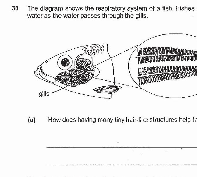
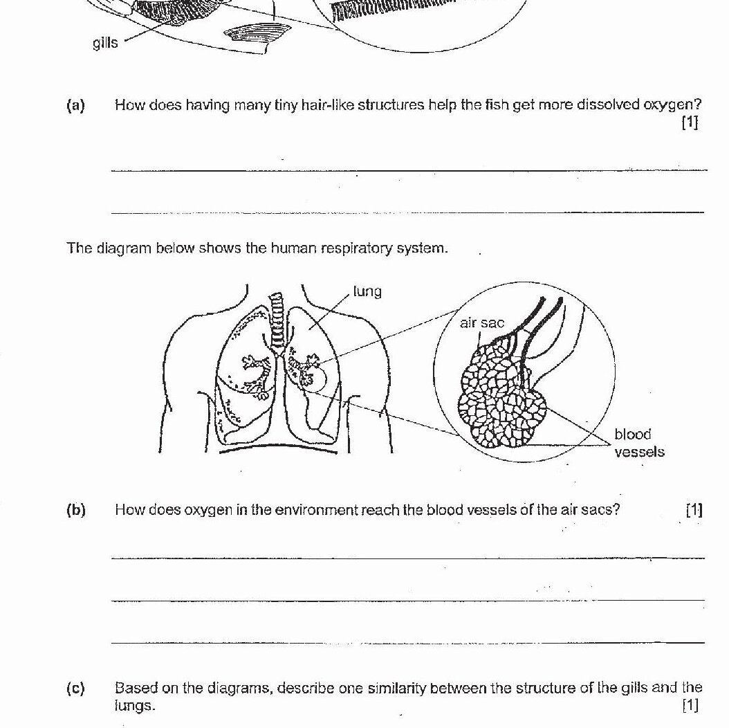
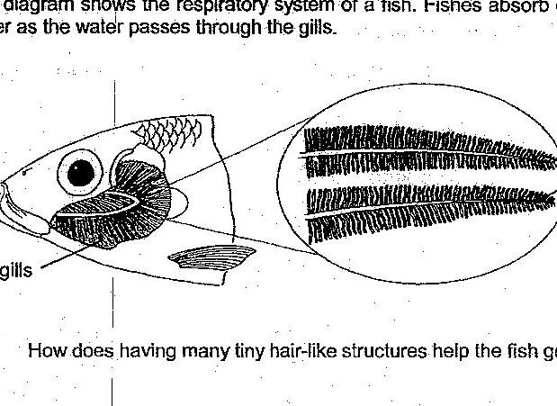
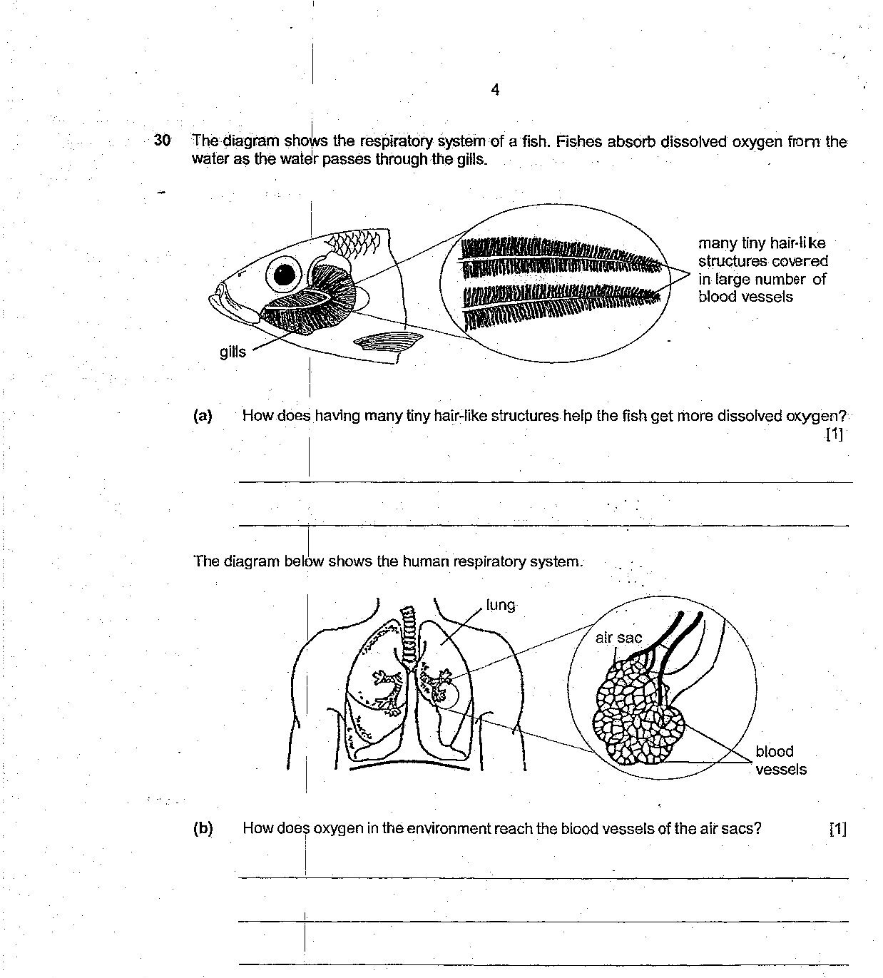
How does having many tiny hair-like structures help the fish get more dissolved oxygen?
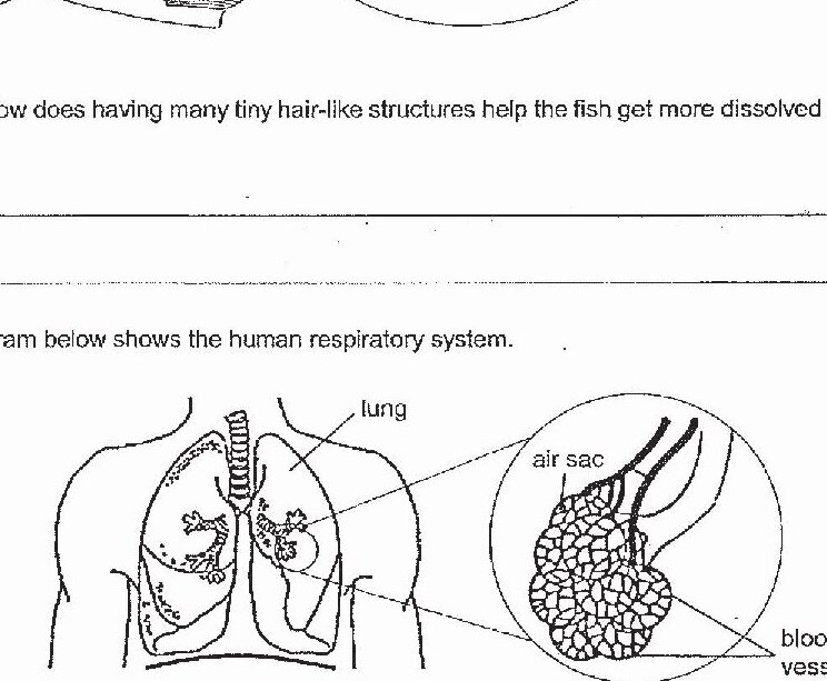
How does oxygen in the environment reach the blood vessels of the air sacs?
Based on the diagrams, describe one similarity between the structure of the gills and the lungs.
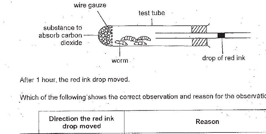
Gabriel set up the apparatus as shown below. In the set-up, the drop of red ink prevents air from entering the test tube. After 1 hour, the red ink drop moved. Which of the following shows the correct observation and reason for the observation?
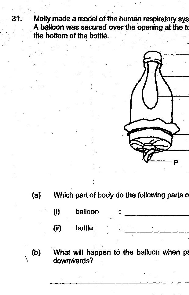
Which part of body do the following parts of the model represent? balloon
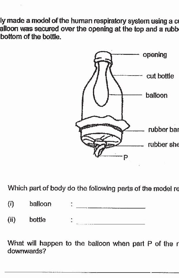
Which part of body do the following parts of the model represent? bottle
What will happen to the balloon when part P of the rubber sheet was pulled downwards?
State one difference between the composition of air entering the lungs and the air leaving the lungs in the human respiratory system.
How does having many tiny hair-like structures help the fish get more dissolved oxygen?
How does oxygen in the environment reach the blood vessels of the air sacs?
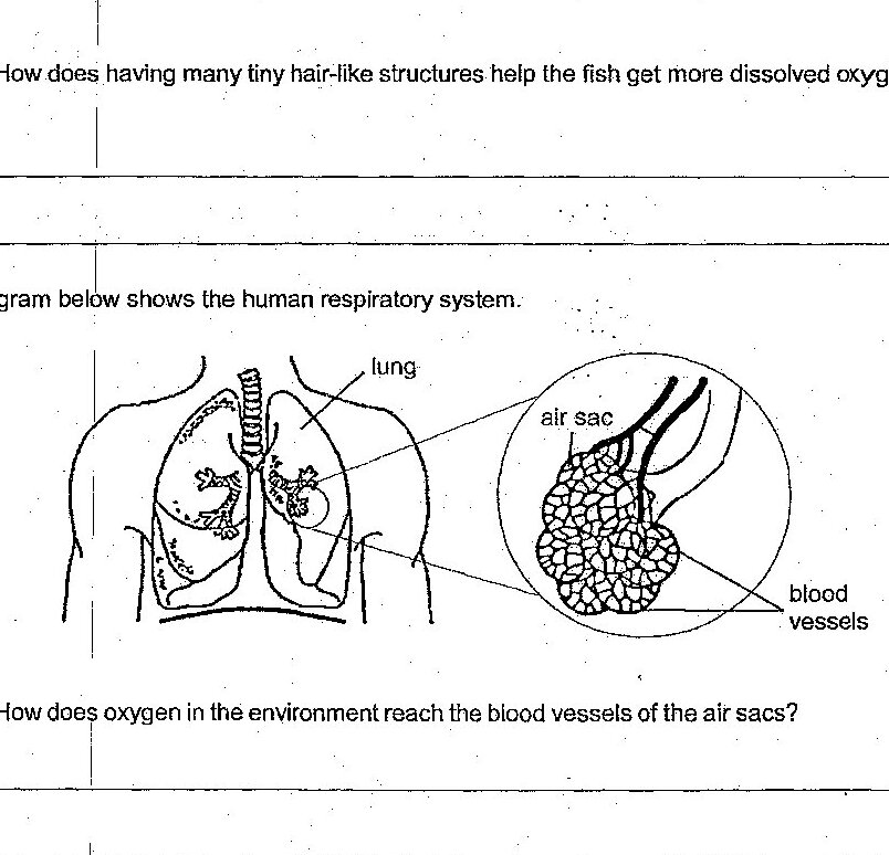
Based on the diagrams, describe one similarity between the structure of the gills and the lungs.
State one difference between the composition of air entering the lungs and the air leaving the lungs in the human respiratory system.
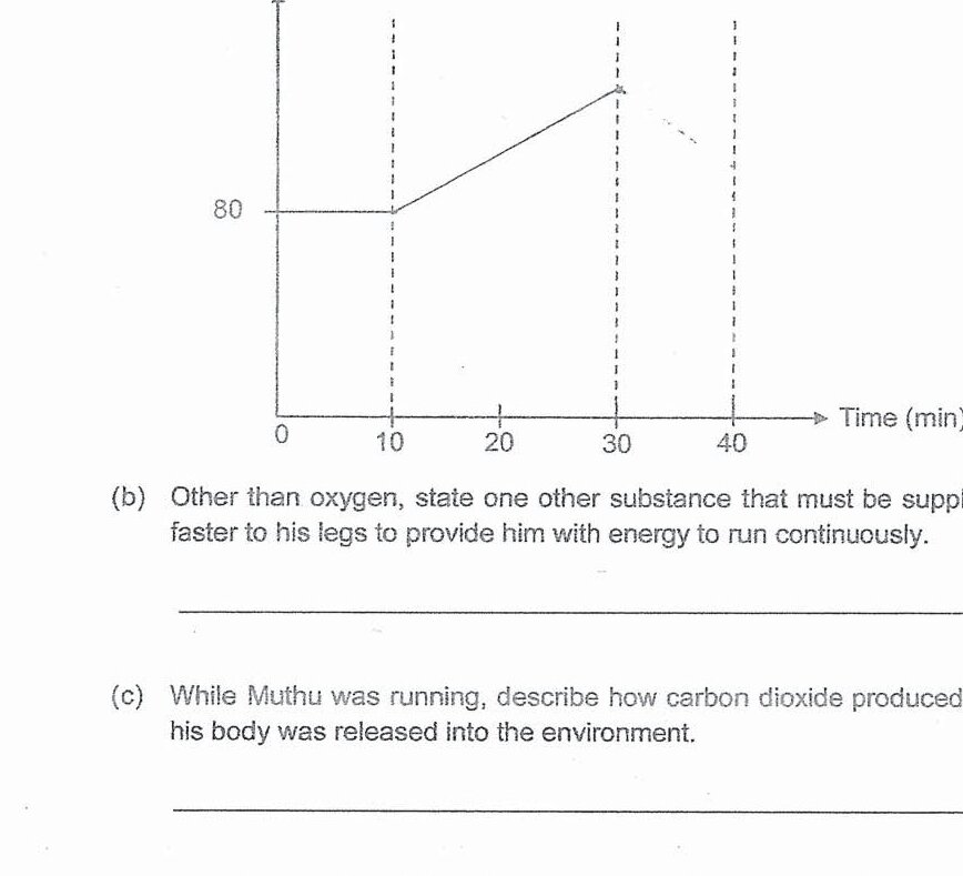
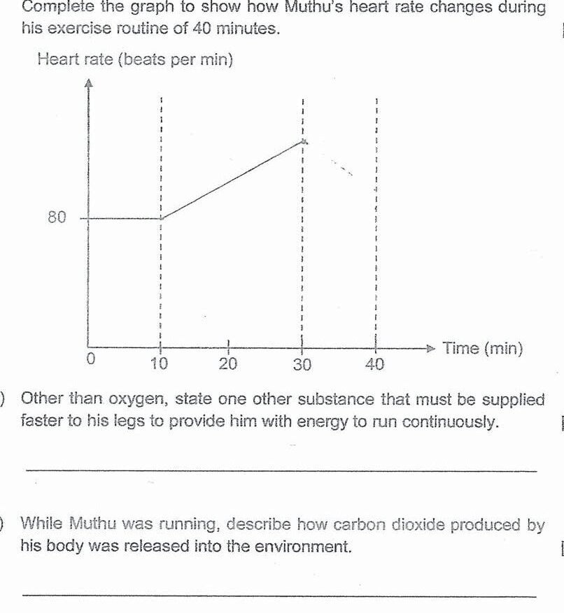
While Muthu was running, describe how carbon dioxide produced by his body was released into the environment.
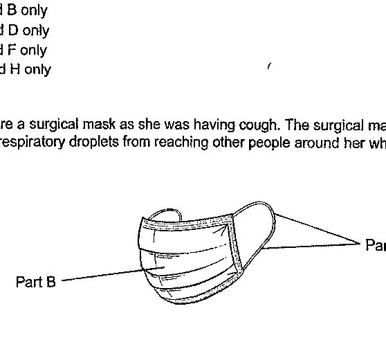
Mrs Woo wore a surgical mask as she was having cough. The surgical mask helps to prevent her respiratory droplets from reaching other people around her when she coughs. What are the properties of the materials used to make parts A and B so that the surgical mask can perform its function?
While Muthu was running, describe how carbon dioxide produced by his body was released into the environment.
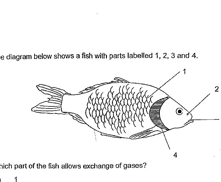
Which part of the fish allows exchange of gases?
Describe how oxygen in the environment reaches the muscles.

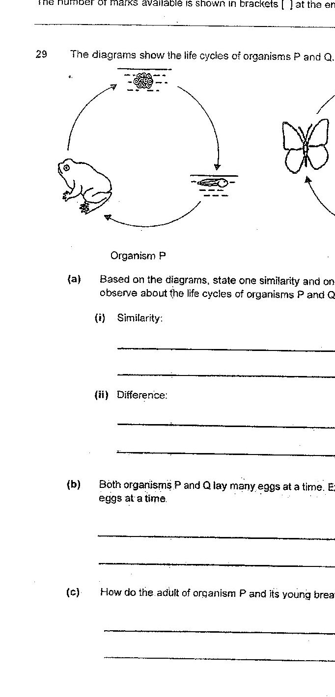
How do the adult of organism P and its young breathe in water?
Describe the change in Mr Tan's breathing rate after getting the bacterial infection and explain why.
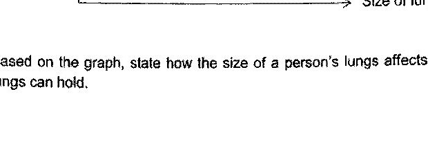
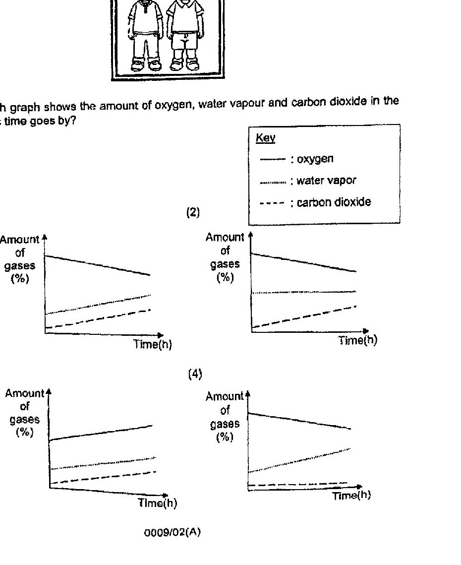
A few people were trapped in a lift during a blackout which caused the fan in the lift to stop working. Which of the following shows the correct changes in the amount of the components of air inside the lift after one hour? A - Amount of oxygen decreases B - Amount of water vapour increases C - Amount of carbon dioxide remains unchanged
Two boys were trapped in a lift for several hours. Which graph shows the amount of oxygen, water vapour and carbon dioxide in the lift as time goes by?
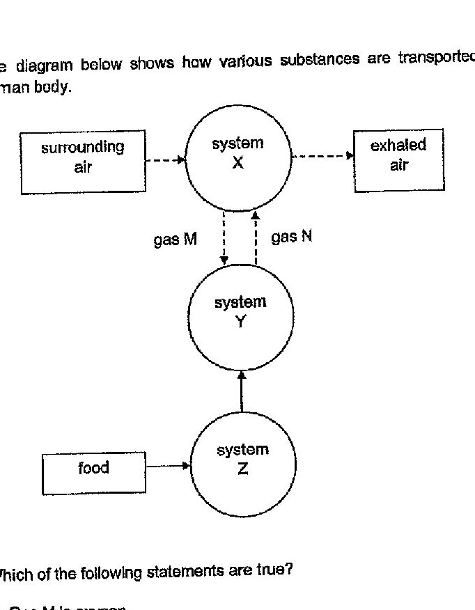
The diagram below shows how various substances are transported in the human body. Which of the following statements are true? A: Gas M is oxygen. B: System Y is involved in gaseous exchange. C: System Z is involved in absorption of simple substances.
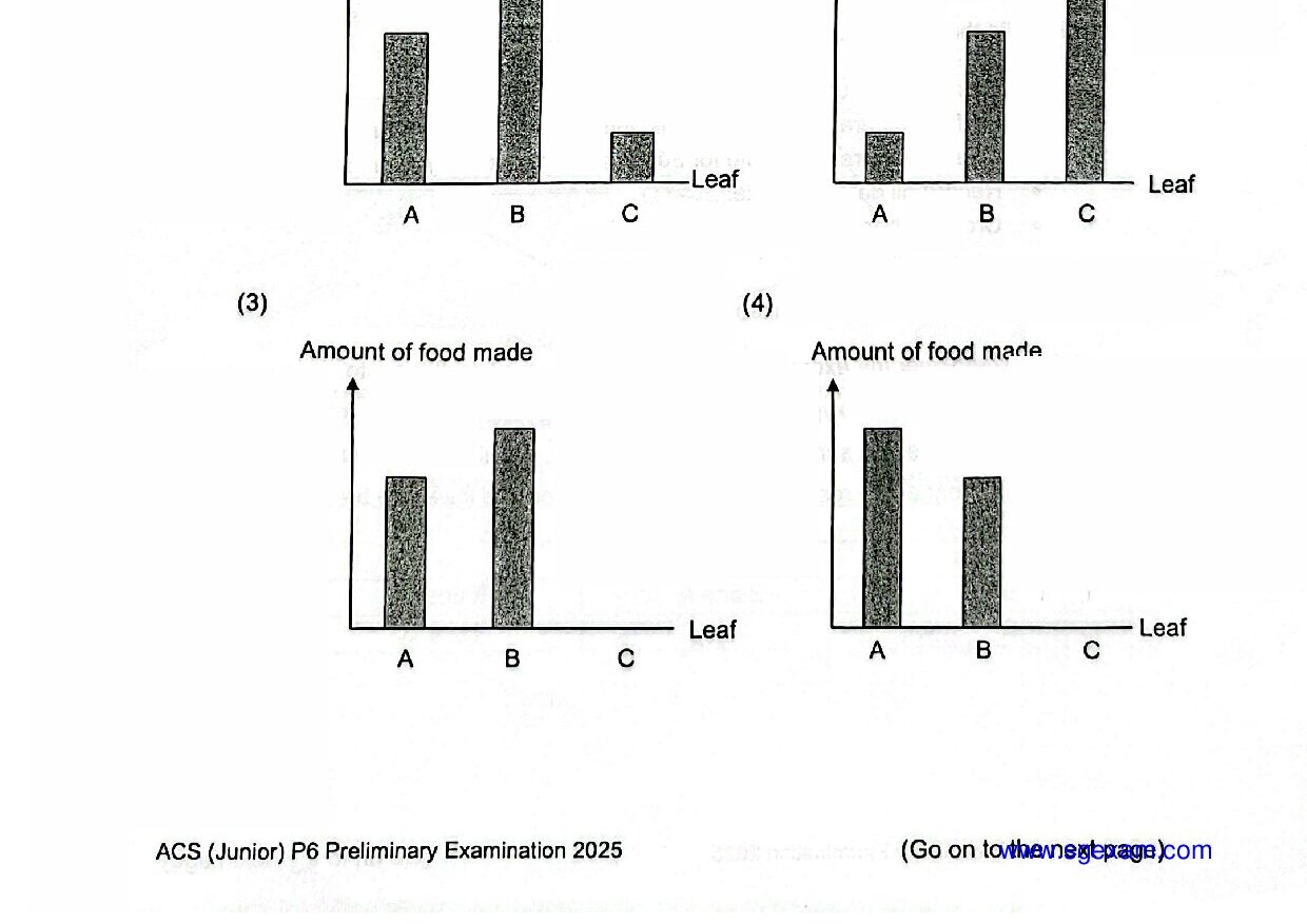
A group of students carried out an investigation to test their hypothesis. They did the following: Rest for 2 minutes and record initial breathing rate. Perform different exercises (walk, jump and run) for 1 minute each. Count their breathing rate for 30 seconds immediately after each exercise. Record all data in a table. Create a bar graph to compare results. Which hypothesis is most suitable for this investigation?
State all the organs in the human respiratory system.
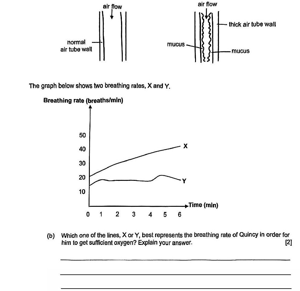
Which one of the lines, X or Y, best represents the breathing rate of Quincy in order for him to get sufficient oxygen? Explain your answer.
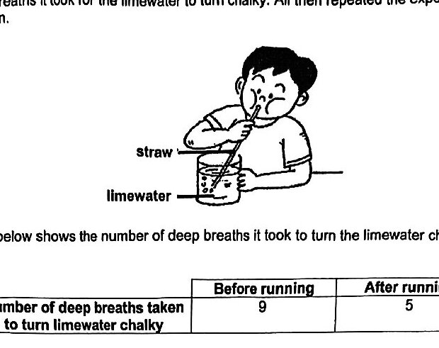
Based on the results above, what can Ali conclude about the amount of carbon dioxide present in exhaled air after running?
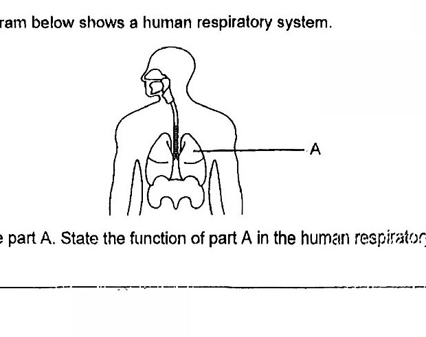
Name part A. State the function of part A in the human respiratory system.
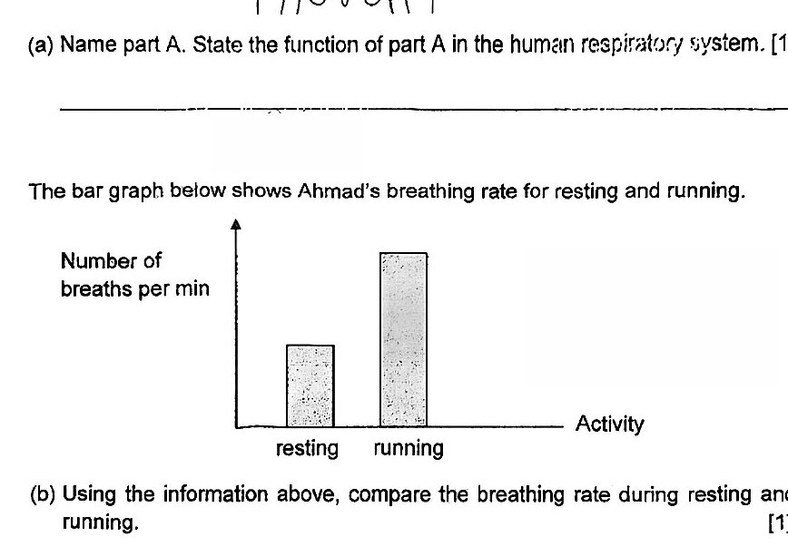
Using the information above, compare the breathing rate during resting and running.
State the function of the human respiratory system. Include in your answer the gases involved.
Past year papers cover the full exam — browse by subject below.
View All Papers ›