Digestive System
Digestive System - Primary 6 PSLE Science Study Notes
Key Concepts
What is the Digestive System?
- The digestive system is a group of organs working together to break down food into smaller particles that can be absorbed by the body
- Food must be broken down because the body cannot use large food particles directly
- The nutrients from digested food provide energy and materials for growth and repair
- There are two types of digestion:
- Physical digestion: Breaking down food into smaller pieces (no new substances formed)
- Chemical digestion: Breaking down food using digestive juices (new substances formed)
The Journey of Food
- Food travels through the digestive system in one direction: mouth → oesophagus (gullet) → stomach → small intestine → large intestine → rectum → anus
- This journey takes approximately 24-72 hours depending on the type of food eaten
- Not all food can be digested - undigested food becomes faeces
Why Do We Need Digestion?
- Large food molecules cannot pass through the walls of the small intestine into the blood
- Digestion breaks down large molecules into small, soluble molecules
- Only small, soluble molecules can be absorbed into the bloodstream
- The blood then transports these nutrients to all parts of the body
Important Definitions
Digestive System: A system of organs that work together to break down food into simpler substances that can be absorbed by the body.
Physical Digestion: The breakdown of food into smaller pieces by physical means (such as chewing or churning) without changing the food chemically. No new substances are formed.
Chemical Digestion: The breakdown of food into simpler substances using digestive juices. New substances are formed during this process.
Digestive Juices: Liquids produced by organs in the digestive system that contain chemicals which break down food during chemical digestion.
Digestion: The process of breaking down large food particles into smaller, simpler and soluble substances that can be absorbed by the body.
Absorption: The process by which digested food (small, soluble nutrients) passes through the wall of the small intestine into the bloodstream.
Peristalsis: The wave-like contractions of muscles in the digestive system that push food along the digestive tract.
Faeces: Undigested food material, dead cells, and bacteria that are removed from the body through the anus.
Rectum: The final section of the large intestine, located just before the anus, which temporarily stores faeces before they are expelled from the body.
Organs of the Digestive System and Their Functions
1. Mouth and Teeth
Structure:
- Contains teeth, tongue, and salivary glands
- Teeth are hard structures made for breaking down food
Functions:
- Teeth: Bite and chew food to break it into smaller pieces (physical digestion)
- Tongue: Mixes food with saliva and helps push food to the back of the mouth for swallowing
- Saliva: Moistens food and begins chemical digestion of starch
- Makes food easier to swallow
Types of Digestion:
- Physical digestion: Chewing with teeth
- Chemical digestion: Saliva breaks down starch
2. Gullet (Oesophagus)
Structure:
- A long, muscular tube connecting the mouth to the stomach
- Approximately 25 cm long
Functions:
- Transports food from the mouth to the stomach
- Muscles in the wall contract and relax (peristalsis) to push food down
- You can swallow even when upside down because of peristalsis!
Types of Digestion:
- No digestion occurs here - it’s a transport tube only
3. Stomach
Structure:
- A J-shaped muscular bag
- Has thick, muscular walls
- Can expand to hold food
Functions:
- Physical digestion: Muscular walls contract and relax to churn food, mixing it with digestive juices
- Chemical digestion: Produces digestive juices (gastric juice) that break down food, especially proteins
- Stores food temporarily (2-4 hours)
- Kills harmful bacteria in food with stomach acid
Types of Digestion:
- Physical digestion: Churning action
- Chemical digestion: Digestive juices break down proteins
4. Small Intestine
Structure:
- A long, coiled tube (approximately 6 meters)
- Inner wall has many finger-like projections called villi (singular: villus)
- Villi increase the surface area for absorption
Functions:
- Chemical digestion: Completes the digestion of food using digestive juices
- Absorption: Digested food (small, soluble nutrients) passes through the walls into the bloodstream
- Most digestion and absorption happens here
- The long length and villi provide a large surface area for maximum absorption
Types of Digestion:
- Chemical digestion: Final breakdown of all food types
- Main site of absorption
5. Large Intestine
Structure:
- A wider but shorter tube (approximately 1.5 meters)
- Surrounds the small intestine
Functions:
- Absorbs water from undigested food
- Temporarily stores faeces (undigested food waste)
- No digestion occurs here
Types of Digestion:
- No digestion - only water absorption
6. Rectum
Structure:
- The final section of the large intestine, located just before the anus
- A short, muscular chamber that connects the large intestine to the anus
Functions:
- Temporarily stores faeces until they are ready to be expelled from the body
- Signals to the body when faeces are ready to be expelled (creates the feeling of needing to use the toilet)
Types of Digestion:
- No digestion - only temporary storage of waste
7. Anus
Structure:
- The opening at the end of the digestive system (at the end of the rectum)
Functions:
- Allows faeces (undigested food) to leave the body
- This process is called egestion or defecation
Types of Digestion:
- No digestion - only removal of waste
The Complete Digestion Process
Step-by-Step Journey of Food
Step 1: Mouth (Physical and Chemical Digestion Begin)
- Teeth bite and chew food into smaller pieces (physical digestion)
- Tongue mixes food with saliva
- Saliva moistens food and begins breaking down starch (chemical digestion)
- Food is formed into a soft ball shape for swallowing
Step 2: Gullet (Transport Only)
- Food is swallowed and enters the gullet
- Muscles in the gullet wall contract and relax in waves (peristalsis)
- Food is pushed down to the stomach (takes a few seconds)
Step 3: Stomach (Physical and Chemical Digestion Continue)
- Muscular walls churn and mix food (physical digestion)
- Digestive juices are added to food (chemical digestion)
- Food becomes a thick liquid mixture
- Proteins begin to be broken down
- Food stays in stomach for 2-4 hours
Step 4: Small Intestine (Chemical Digestion Completed and Absorption)
- More digestive juices are added
- All remaining food is broken down into small, soluble substances (chemical digestion)
- Digested food (nutrients) passes through the intestine walls into the bloodstream (absorption)
- Blood carries nutrients to all parts of the body
- Food stays here for 3-5 hours
Step 5: Large Intestine (Water Absorption)
- Undigested food enters the large intestine
- Water is absorbed from the undigested food
- Undigested food becomes more solid and forms faeces
- Food stays here for 10-24 hours or longer
Step 6: Rectum (Temporary Storage of Faeces)
- Faeces passes from the large intestine into the rectum
- The rectum stores faeces temporarily until they are ready to be expelled
- When the rectum is full, the body signals the need to defecate
Step 7: Anus (Removal of Waste)
- Faeces (undigested food) leaves the body through the anus
- This is called egestion or defecation
Chemical Digestion
What Happens in Chemical Digestion?
- Digestive juices are produced by organs such as the mouth (saliva), stomach, and small intestine
- Digestive juices break down large food molecules into smaller, soluble molecules that can be absorbed into the blood
- Chemical digestion occurs mainly in the stomach and small intestine
- Digestive juices work best at body temperature (37°C) — too hot or too cold reduces their effectiveness
Note: Specific enzyme names (amylase, protease, lipase) and their individual substrates are not required knowledge for PSLE. The term “digestive juices” is sufficient.
Worked Examples
Example 1: Identifying Types of Digestion
Question: Ali eats a sandwich. Identify whether physical or chemical digestion occurs at each stage.
| Location | Process | Type of Digestion |
|---|---|---|
| Mouth | Teeth chew the sandwich | Physical digestion |
| Mouth | Saliva mixes with sandwich | Chemical digestion |
| Gullet | Sandwich moves down | No digestion (transport only) |
| Stomach | Stomach walls churn sandwich | Physical digestion |
| Stomach | Digestive juices mix with sandwich | Chemical digestion |
| Small intestine | Digestive juices break down sandwich | Chemical digestion |
| Large intestine | Water absorbed from undigested food | No digestion (absorption only) |
Key Points:
- Physical digestion = Breaking into smaller pieces (chewing, churning)
- Chemical digestion = Using digestive juices to break down food
- Some organs do both types, some do neither
Example 2: Explaining Absorption
Question: Explain why digestion must occur before absorption can take place in the small intestine.
Model Answer (Step-by-step):
-
State what needs to be absorbed:
- Nutrients from food need to be absorbed into the bloodstream.
-
Explain the problem with large particles:
- Large food particles cannot pass through the walls of the small intestine.
-
Explain what digestion does:
- Digestion breaks down large food particles into small, soluble substances.
-
Connect to absorption:
- Only small, soluble substances can pass through the walls of the small intestine into the bloodstream.
-
Conclude:
- Therefore, digestion must occur first to make food particles small enough to be absorbed.
Complete Answer: “Digestion must occur before absorption because large food particles cannot pass through the walls of the small intestine. Digestion breaks down large food particles into small, soluble substances. Only these small, soluble substances can pass through the intestinal walls into the bloodstream to be transported to different parts of the body.”
Example 3: Functions of the Small Intestine
Question: The small intestine is very long and has many finger-like structures called villi on its inner walls. Explain how these features help the small intestine carry out its function.
Model Answer (Step-by-step):
-
State the main functions:
- The small intestine’s main functions are digestion and absorption.
-
Explain the long length:
- The small intestine is very long (about 6 meters), which provides a large surface area for digestion and absorption to occur.
- Food stays in the small intestine for a longer time, allowing complete digestion and absorption.
-
Explain the villi:
- The inner walls have many finger-like structures called villi.
- These villi greatly increase the surface area of the small intestine.
- A larger surface area allows more digested food to be absorbed into the bloodstream at the same time.
-
Conclude:
- These features make absorption more efficient.
Complete Answer: “The small intestine is very long (about 6 meters), which provides a large surface area and gives more time for digestion and absorption to occur. The villi are finger-like structures on the inner walls that greatly increase the surface area. This larger surface area allows more digested food to be absorbed into the bloodstream efficiently.”
Common Mistakes to Avoid
Mistake 1: Confusing Small and Large Intestines
❌ Wrong: “The large intestine is longer than the small intestine.” ✅ Correct: “The small intestine is longer (about 6m) but narrower. The large intestine is shorter (about 1.5m) but wider.”
Why it matters: Students mix up the names with the actual size. Remember: “small” and “large” refer to WIDTH, not length.
Mistake 2: Saying Digestion Happens Everywhere
❌ Wrong: “Digestion occurs in the gullet and large intestine.” ✅ Correct: “Digestion occurs in the mouth, stomach, and small intestine. The gullet only transports food. The large intestine only absorbs water.”
Why it matters: Not all organs digest food - some have other functions.
Mistake 3: Confusing Digestion with Absorption
❌ Wrong: “The stomach absorbs nutrients into the blood.” ✅ Correct: “The stomach digests food. The small intestine absorbs digested food (nutrients) into the blood.”
Why it matters: Digestion (breaking down) and absorption (taking in nutrients) are different processes. Absorption mainly happens in the small intestine.
Mistake 4: Incomplete Explanations for Physical vs Chemical Digestion
❌ Wrong: “Chewing is physical digestion because it breaks down food.” ✅ Correct: “Chewing is physical digestion because it breaks down food into smaller pieces without changing the food chemically. No new substances are formed.”
Why it matters: You need to mention that no new substances are formed in physical digestion to get full marks.
Mistake 5: Forgetting That Food Must Be Soluble
❌ Wrong: “Digestion makes food smaller so it can be absorbed.” ✅ Correct: “Digestion breaks down food into small AND soluble substances so they can be absorbed.”
Why it matters: Both “small” and “soluble” are keywords. Food must be soluble to dissolve and pass through the intestine walls.
Mistake 6: Wrong Order of Organs
❌ Wrong: “Food goes from mouth → stomach → gullet → small intestine.” ✅ Correct: “Food goes from mouth → oesophagus (gullet) → stomach → small intestine → large intestine → rectum → anus.”
Why it matters: The sequence is important and commonly tested. From 2026, the rectum is a named organ in the digestive pathway — do not skip it.
Mistake 7: Saying Faeces Contains Digested Food
❌ Wrong: “Faeces contains digested food that leaves the body.” ✅ Correct: “Faeces contains undigested food that leaves the body through the anus.”
Why it matters: Digested food is absorbed into the blood - it doesn’t leave as faeces.
Mistake 8: Incomplete Function Descriptions
❌ Wrong: “The stomach digests food.” ✅ Correct: “The stomach carries out physical digestion by churning food and chemical digestion using digestive juices.”
Why it matters: For function questions, mention both HOW (physical/chemical) and WHAT (churning/digestive juices).
Mistake 9: Misreading ‘Leaving’ vs ‘Entering’ in Diagram Questions
❌ Wrong: Identifying the organ by the food entering it, leading to the wrong organ being named. ✅ Correct: Read the question carefully — if it asks about food ‘leaving’ an organ, that food has already been processed there. Track whether food is entering or exiting to correctly name the organ.
Why it matters: Exam diagrams often test whether you understand the state of food at each stage. The keyword ‘leaving’ means the food has already been through that organ’s processes.
Exam Tips
Keywords to Include in Answers
For Digestion Questions:
- “Physical digestion” and “chemical digestion” (specify which type)
- “Break down food into smaller pieces” (physical)
- “Break down food using digestive juices” (chemical)
- “No new substances formed” (physical) or “New substances formed” (chemical)
- “Small and soluble substances”
For Absorption Questions:
- “Small and soluble” (both words needed!)
- “Pass through the walls of the small intestine”
- “Into the bloodstream”
- “Transported to different parts of the body”
For Function Questions:
- Always mention BOTH physical and chemical digestion if the organ does both
- State what specific action occurs (e.g., “chewing,” “churning,” “mixing with digestive juices”)
Mark-Earning Phrases
High-value phrases for different question types:
-
Why must food be digested?
- “Large food particles cannot pass through the walls of the small intestine”
- “Must be broken down into small and soluble substances”
- “So they can be absorbed into the bloodstream”
-
Difference between physical and chemical digestion:
- Physical: “Breaking food into smaller pieces without chemical change / no new substances formed”
- Chemical: “Breaking down food using digestive juices / new substances formed”
-
Function of small intestine:
- “Completes chemical digestion of food”
- “Absorbs digested food into the bloodstream”
- “Has villi to increase surface area for absorption”
-
Why is the small intestine long?
- “Provides large surface area”
- “Allows more time for digestion and absorption”
- “Allows more digested food to be absorbed”
-
Function of large intestine:
- “Absorbs water from undigested food”
- “Stores faeces temporarily”
- “No digestion occurs”
Answering Strategy
For “State” questions (1 mark each):
- Give a brief, direct answer
- Example: “State one function of the stomach.” → “To digest food” OR “To churn food”
For “Explain” questions (2-3 marks):
- Use the Point-Explain-Link structure
- Give a reason using “because,” “so that,” or “this allows”
- Example: “Explain why the small intestine has villi.”
- Point: The villi increase the surface area of the small intestine.
- Link: This allows more digested food to be absorbed into the bloodstream.
For “Compare” or “Difference” questions:
- Make sure you mention BOTH things being compared
- Example: “State one difference between physical and chemical digestion.”
- “Physical digestion breaks food into smaller pieces while chemical digestion uses digestive juices to break down food.” ✓
- NOT: “Physical digestion breaks food into smaller pieces.” ✗ (incomplete - didn’t mention chemical)
For Function Questions:
- If an organ does multiple things, mention all of them
- Stomach does: physical digestion (churning) + chemical digestion (digestive juices)
- Small intestine does: chemical digestion + absorption
For diagram identification questions: Focus on whether the question says ‘leaving’ or ‘entering’ an organ. Food ‘leaving’ an organ has already been processed there. This helps you correctly identify which organ is shown and what has happened to the food at that point.
Drawing Diagrams
If asked to draw the digestive system:
- Draw organs in the correct order from top to bottom
- Make the small intestine longer and more coiled than the large intestine
- Make the large intestine wider than the small intestine
- Label all parts clearly with arrows pointing to each organ
- Don’t forget the anus!
- Don’t forget the rectum (between large intestine and anus)!
Common labeling errors to avoid:
- Don’t label the gullet as “food pipe” (use “gullet” or “oesophagus”)
- Don’t label the large intestine as “colon”
- Don’t leave out any organs (including the rectum!)
Quick Summary
Essential Points for Revision:
✓ Order of organs: Mouth → Oesophagus (Gullet) → Stomach → Small intestine → Large intestine → Rectum → Anus
✓ Two types of digestion: Physical (breaking into smaller pieces, no new substances) and Chemical (using digestive juices, new substances formed)
✓ Mouth: Physical digestion (teeth chew) + Chemical digestion (saliva breaks down starch)
✓ Oesophagus/Gullet: Transports food using peristalsis (wave-like muscle contractions) - NO digestion occurs
✓ Stomach: Physical digestion (churning) + Chemical digestion (digestive juices break down proteins) - stores food for 2-4 hours
✓ Small intestine: Completes chemical digestion + Absorbs digested food into bloodstream - LONGEST organ (6m), has villi to increase surface area
✓ Large intestine: Absorbs water from undigested food - NO digestion, wider but shorter (1.5m)
✓ Rectum: Temporarily stores faeces before they leave the body
✓ Anus: Removes faeces (undigested food) from the body
✓ Why digest food: Large particles cannot pass through intestine walls → Must break down into small AND soluble substances → Then can be absorbed into blood
✓ Absorption happens mainly in: Small intestine (digested food passes through walls into bloodstream)
✓ Remember: Small intestine is LONGER but NARROWER; Large intestine is SHORTER but WIDER
✓ Faeces contains: Undigested food (digested food is absorbed into blood, not removed as faeces)
Final Check Before Exam:
- Can you name all 7 parts of the digestive system in order (including rectum)?
- Can you explain the difference between physical and chemical digestion?
- Do you know which organs do physical, chemical, or both types of digestion?
- Can you explain why food must be “small and soluble” to be absorbed?
- Can you describe what happens in each organ?
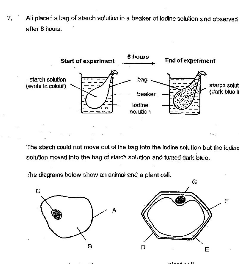
Which parts of the cells have similar function as that of the bag in Ali's experiment?
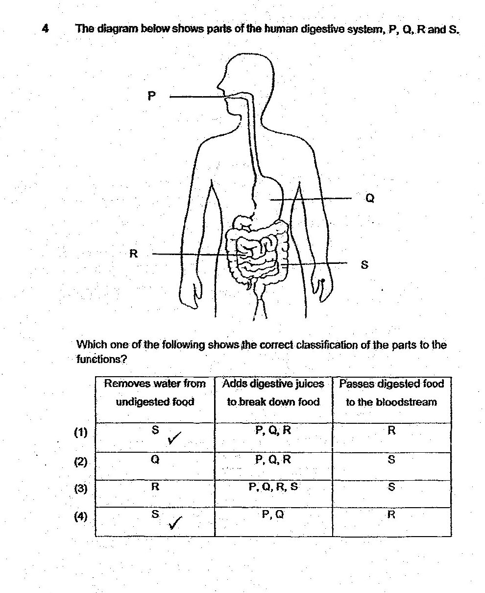
The diagram below shows parts of the human digestive system, P, Q, R and S. Which one of the following shows the correct classification of the parts to the functions?
Based on Jordan's set-up above, complete the circuit diagram of circuit Q. In the space below, draw a light bulb, buzzer and wires so that the light bulb will switch on and the buzzer will sound when someone steps onto set-up P. Switch P is used to represent set-up P. Use (B) to represent the buzzer.
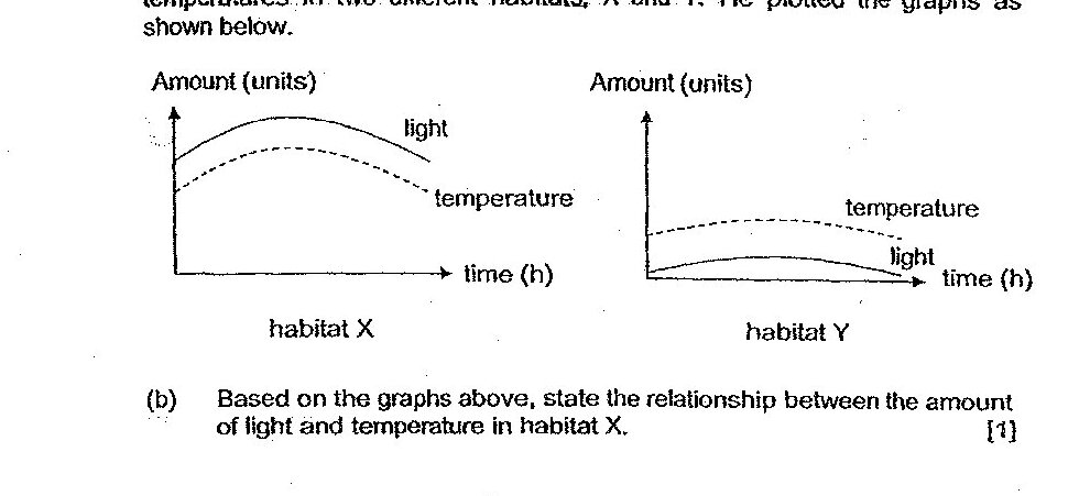
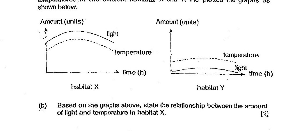
Based on the information in the table and graphs, compare the amount of moisture that is most likely found in habitats X and Y.
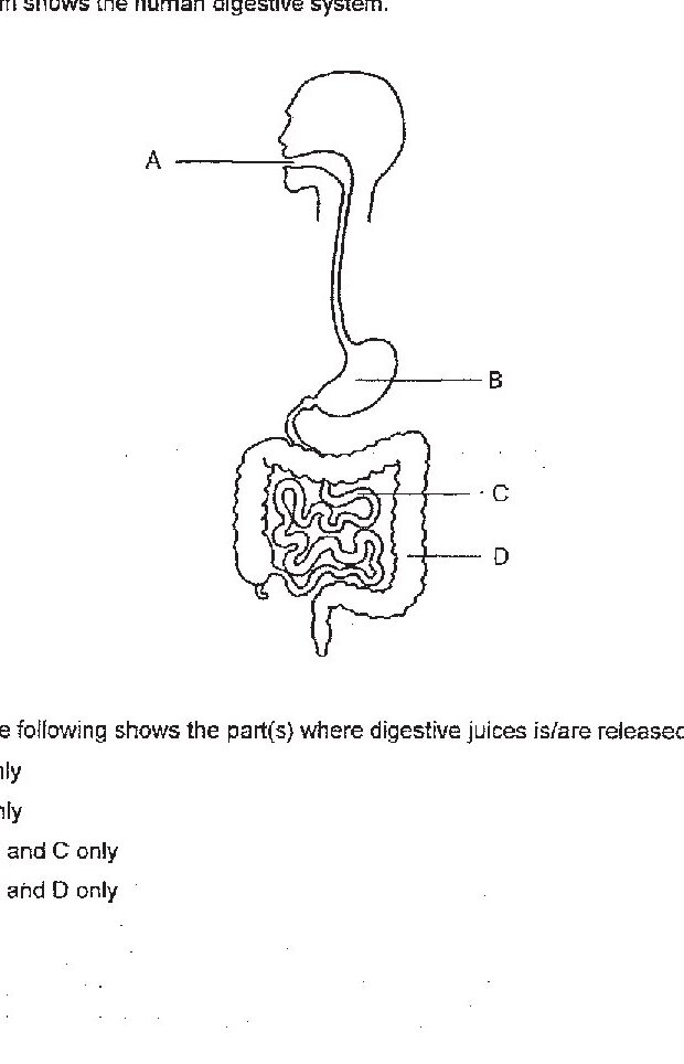
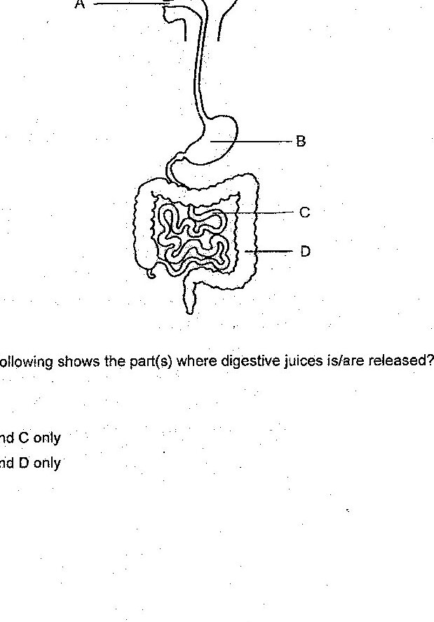
Which of the following shows the part(s) where digestive juices is/are released?
Explain why the mass of the food pieces in all three beakers decreased.
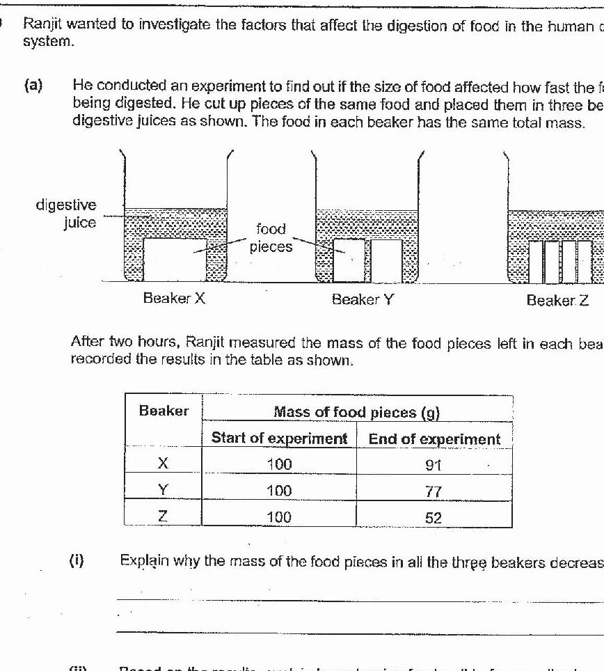
Based on the results, explain how chewing food well before swallowing can help for food to be digested faster.

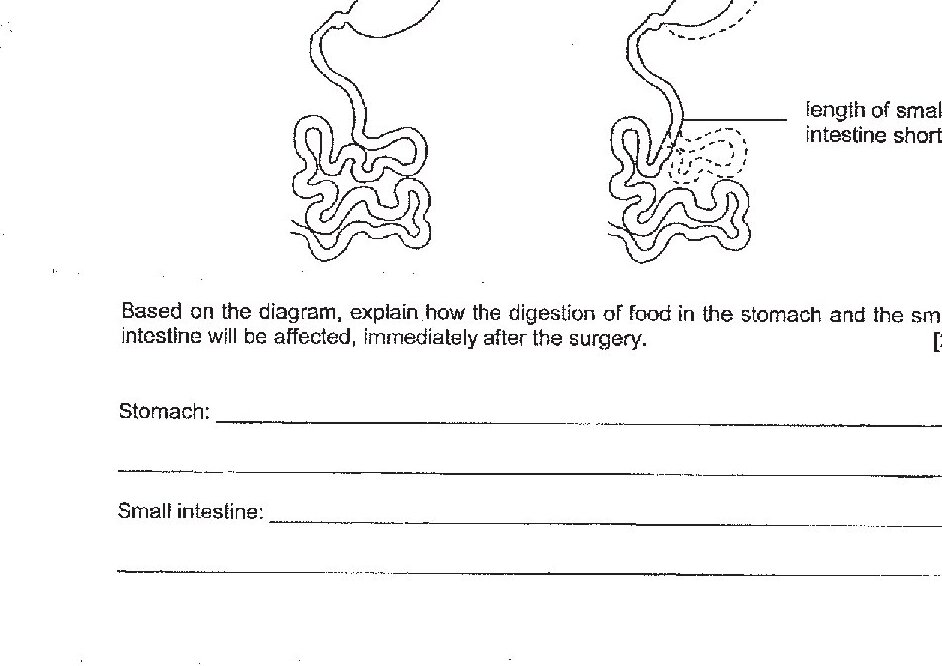
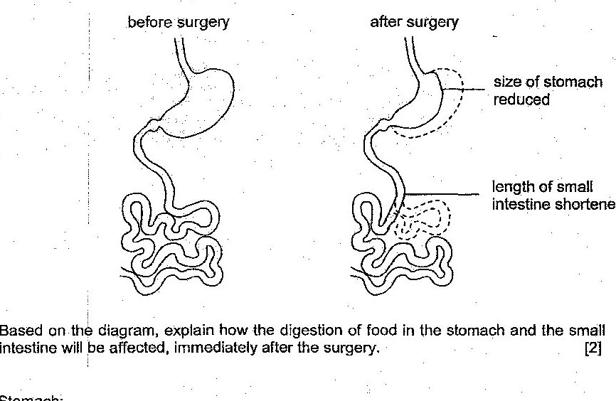
Based on the diagram, explain how the digestion of food in the stomach and the small intestine will be affected, immediately after the surgery.
From which of the following organs will water be absorbed into the bloodstream?
Study the flow chart below. Which of the following correctly represents systems X, Y and Z?
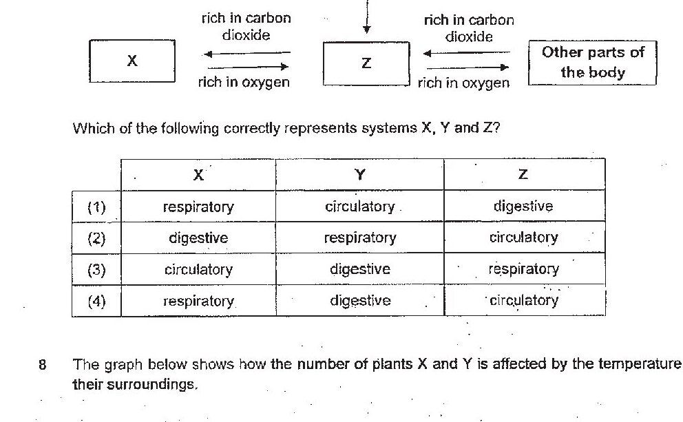
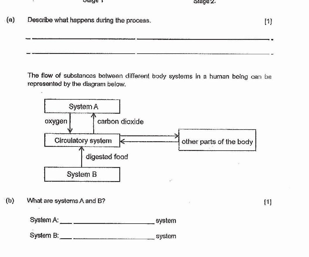
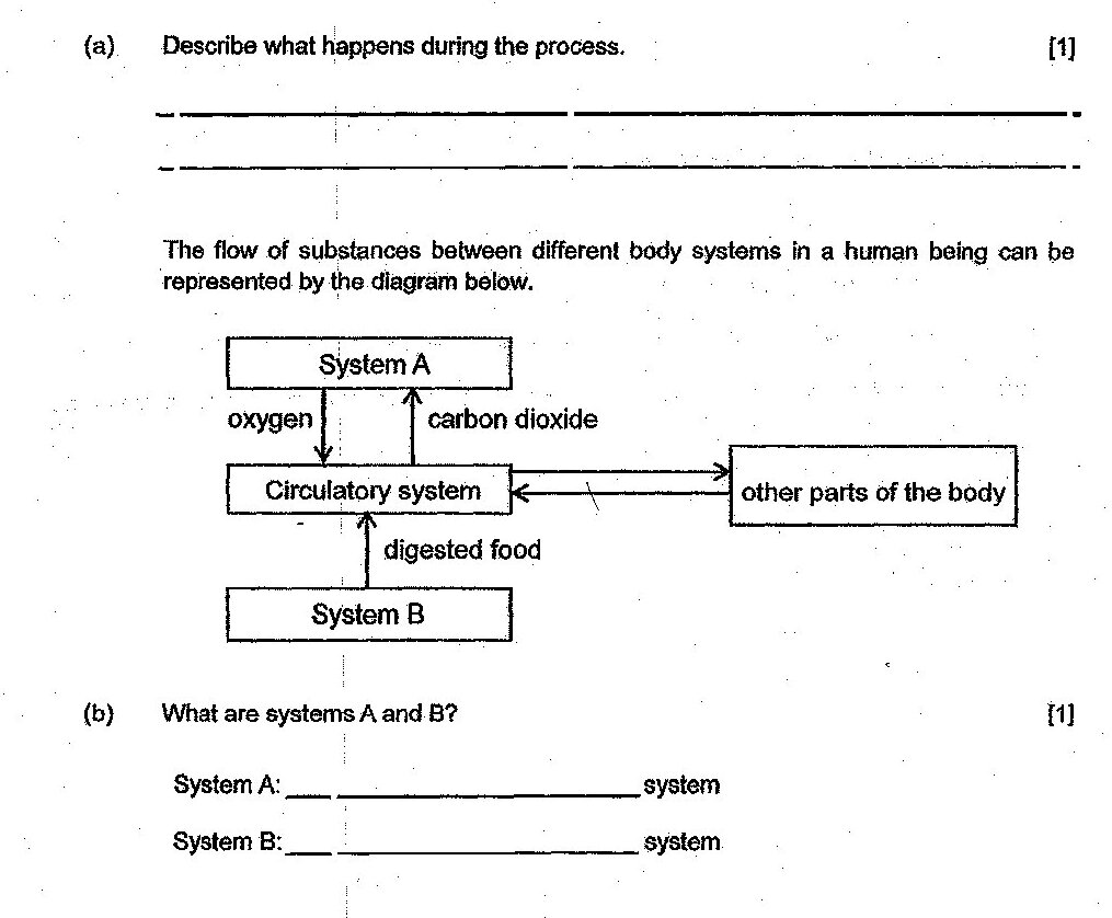
What are systems A and B?
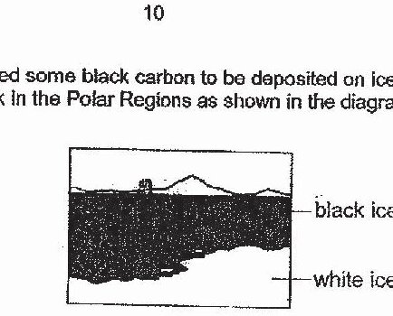
Based on the above results and information, explain how this would reduce the population size of Animal P staying in the Polar Regions.
Which of the following shows the part(s) where digestive juices is/are released?
Explain why the mass of the food pieces in all the three beakers decreased.
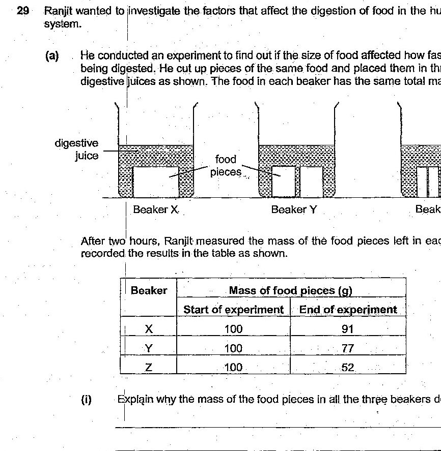
Based on the results, explain how chewing food well before swallowing can help food to be digested faster.
Based on the diagram, explain how the digestion of food in the stomach and the small intestine will be affected, immediately after the surgery.
What are systems A and B?
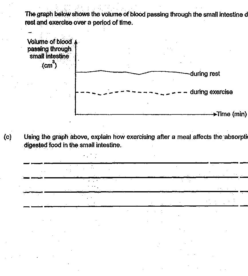
Using the graph above, explain how exercising after a meal affects the absorption of digested food in the small intestine.
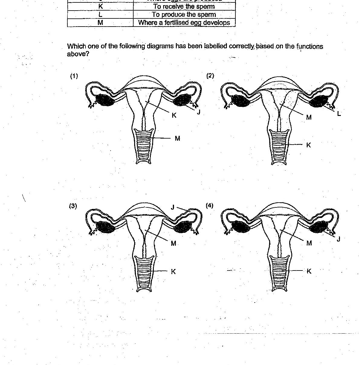
Study the table below. Which one of the following diagrams has been labelled correctly, based on the functions above?
Which of the following correctly describes the functions of the digestive system?
Which combination correctly describes the functions of the digestive system?
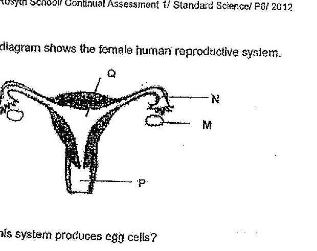
Which part of this system produces egg cells?
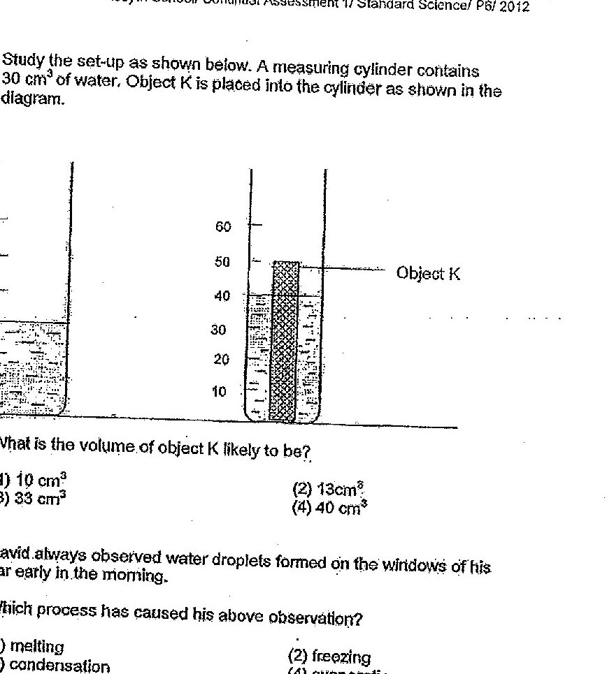
Study the set-up as shown below. A measuring cylinder contains 30 cm³ of water. Object K is placed into the cylinder as shown in the diagram. What is the volume of object K likely to be?
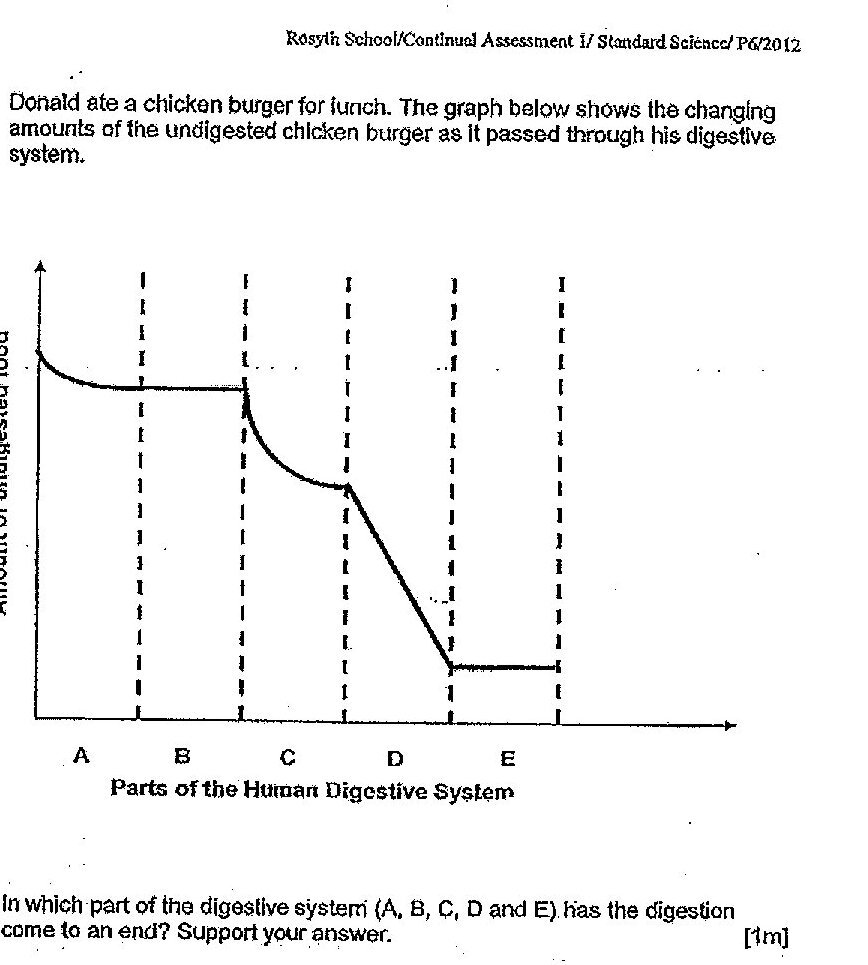
In which part of the digestive system (A, B, C, D and E) has the digestion come to an end? Support your answer.
What happens to the undigested food before it is released from the body?
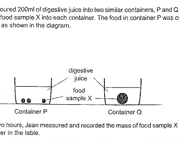
Jean poured 200ml of digestive juice into two similar containers, P and Q. She added 10g of food sample X into each container. The food in container P was cut into small pieces as shown in the diagram. After two hours, Jean measured and recorded the mass of food sample X left in each container in the table. Container | Mass of food left in the container (g) P | 4 Q | 7 (a) What is the changed variable of Jean’s experiment?
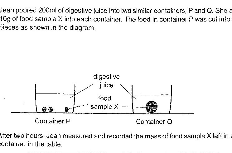
Jean poured 200ml of digestive juice into two similar containers, P and Q. She added 10g of food sample X into each container. The food in container P was cut into small pieces as shown in the diagram. After two hours, Jean measured and recorded the mass of food sample X left in each container in the table. Container | Mass of food left in the container (g) P | 4 Q | 7 (b) What should Jean do to obtain more reliable results?
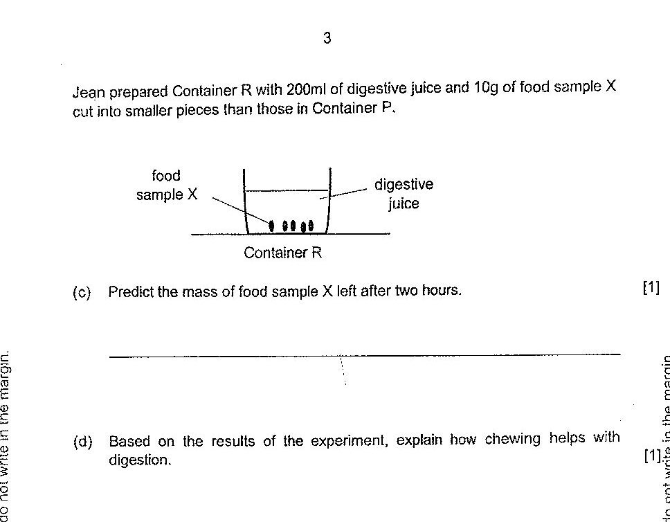
Jean prepared Container R with 200ml of digestive juice and 10g of food sample X cut into smaller pieces than those in Container P. Predict the mass of food sample X left after two hours.
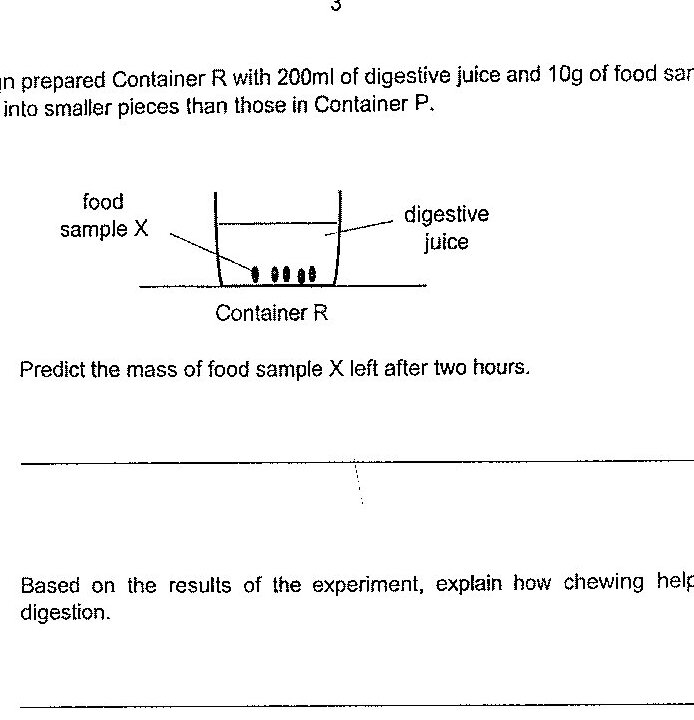
Based on the results of the experiment, explain how chewing helps with digestion.
Past year papers cover the full exam — browse by subject below.
View All Papers ›